If you get called back for additional tests after a mammogram, there’s no need to panic. That advice comes from Jennifer Yanak, the nurse practitioner who manages the high-risk breast clinic at Rush Copley Medical Center.
“That callback doesn’t automatically mean breast cancer,” Yanak says. “You’ll get called back anytime we find something that we want to double-check.” It’s not uncommon to get called back — and, according to the American Cancer Society, less than 1% of women who need further testing are found to have breast cancer.
Screening vs. diagnostic
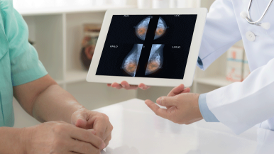
A screening mammogram is part of your annual wellness check. “We know that mammography saves lives,” says Lisa Stempel, MD, division chief of breast imaging at Rush University Medical Center. “Early detection leads to better prognosis: If we find breast cancer at stage 0 or stage 1, the chance of cure is more than 95%.”
A diagnostic radiologist who specializes in breast imaging reads your mammogram and reports the results to you and your provider. The callback rate is highest after the first screening mammogram, since the radiologist doesn’t have older images to compare in order to see whether anything has changed.
If the report after your screening mammogram shows a potentially abnormal finding, you’ll be called back for a diagnostic mammogram. During the diagnostic mammogram, the technologist will focus on the area of concern, such as re-compressing the problem spot to see whether that produces a normal result.
If the screening mammogram appears to show a mass, you may have a handheld ultrasound that gives the diagnostic radiologist who reads your images a closer look at the area. Performed by a specially trained technician, this noninvasive test uses sound waves to take pictures of your breast and provide better images of dense tissue.
You’ll get your diagnostic test report while you wait. If your report after a screening or diagnostic mammogram says that you have a cyst or a benign grouping of calcifications (small deposits of calcium within the breast tissue) and doesn’t advise further tests, you can rest easy, Yanak says. “If the radiologist had a concern, they’d call you back for more imaging or a needle biopsy, which is normally well-tolerated.”
If the radiologist sees an abnormality during your callback testing, your provider will work with you to plan what comes next — whether that’s further testing right away or in a few months. You’ll also be connected with a breast nurse navigator who can answer all your questions about the diagnostic process and accompany you every step of the way if you need to have a needle biopsy.
Personalized medicine based on your risk
The first time you have a screening mammogram at any Rush breast imaging location, you’ll be offered an assessment of your lifetime risk for developing breast cancer, and you’ll also be informed if you have dense breast tissue.
If you choose to be assessed, your imaging technologist will ask questions about your personal and family history and enter your answers into a tablet or computer. Rush’s risk assessment tool then runs the information through a number of scientifically validated risk calculators.
If your lifetime risk is calculated at 20% or more, “you’ll get a phone call from our nurse navigator,” Stempel says. “She’ll explain what the results show, and she’ll offer to refer you to our high-risk clinic.
“This really sets Rush apart. We’re one of the only academic medical centers that ties risk information to a high-risk clinic where patients are managed by experts in breast disease.”
In the high-risk clinic, you’ll meet with specialists — who might include breast surgeons, advanced practice providers and genetic counselors — to create your personalized plan for ongoing regular breast screening and follow-up.
Learn more about our personalized breast cancer screening program and high-risk clinic.
What is supplemental screening for breast cancer?
Because mammography performs less well in women who are high risk or have dense breasts, your plan might include additional screenings added to your yearly mammogram.
Automated breast ultrasound, or ABUS, “is a wonderful technology that helps us find small cancers in dense breast tissue,” Stempel says. “Our ABUS program is finding an additional three to four cancers per thousand that are not seen on the mammogram — and 90% of these cancers are stage 0 and stage 1, when they’re most treatable with less invasive surgery and less need for chemotherapy.”
Since 2009, the American Cancer Society has recommended yearly breast MRI for women who are high risk. Rush now offers fast breast MRI, which requires less than 10 minutes of scan time.
A new offering at Rush University Medical Center is contrast-enhanced mammography. This screening is performed like a regular mammogram, but during the procedure you’ll receive a contrast agent through an IV in your hand or arm. The contrast agent highlights areas of increased blood supply in the breast, which can be an indicator of cancer.
Depending on your unique risk profile, genetic testing and risk-reducing medications could also be among the options you discuss with the high-risk clinic team. They’ll make sure you have a complete understanding of each recommendation, Yanak says. “The choice is always yours — we educate you about your options, and you make an informed decision.”


