When History Misleads — Occult Carcinoma Unmasked by Orbital Biopsy in a Presumed Choriocarcinoma Metastasis
Case report by Maxwell Wilberding, MD, Mohit Uppal, MD, and Kathryn P. Winkler, MD
Introduction
Orbital metastasis is an uncommon but sight-threatening and potentially life-threatening manifestation of systemic malignancy. These metastasis can occurr in up to roughly 3% of cancer patients and frequently producing rapidly progressive proptosis, motility restriction, and optic neuropathy.[1] Breast carcinoma is the single most common source of orbital metastasis, accounting for nearly half of cases, but the orbit can be seeded by carcinomas of the lung, prostate, kidney, thyroid, and neuroendocrine and gestational-trophoblastic origin, among others.[2]
A central diagnostic pitfall is assuming that a new orbital mass, in a patient with a known prior malignancy, is attributed to that cancer. Often the clinical and radiographic picture of orbital metastasis is notoriously nonspecific. Also, the histology of an orbital lesion may be discordant with the presumed—or even the biopsy-proven—primary cancer, therefore materially changing treatment.[1,3] This case describes a woman with a remote history of gestational choriocarcinoma who presented with a fulminant, vision-destroying orbital syndrome radiographically presumed to represent choriocarcinoma metastasis, in whom urgent orbitotomy and biopsy instead revealed a carcinoma of unexpected lineage. It illustrates the limits of clinical–radiographic presumption and the necessity of tissue diagnosis in guiding oncologic care.
Case Presentation
A 49-year-old woman presented to an ophthalmology clinic with a chief complaint of two months of progressive left eyelid swelling, tearing, and gradually decreasing vision in summer of 2026. Her medical history was notable for choriocarcinoma. This was diagnosed in 2020, with the workup revealing a primary pulmonary etiology. She was subsequently treated with chemotherapy and lobectomy and underwent a diagnostic total abdominal hysterectomy/bilateral salpingo-oophorectomy without gynecologic malignancy identified. Following treatment, she was presumed in remission since 2021. However, a loss of medical insurance coverage prevented further follow up for several years. Additional medical history included hypertension, a ruptured intracranial aneurysm complicated by hemorrhagic stroke in 2020 and a prior deep vein thrombosis and pulmonary embolism. She was followed annually by neurosurgery for her aneurysm history but was overdue, with her last interval neuroimaging in 2021.
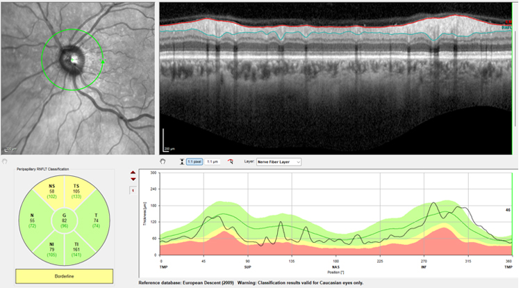
On examination, visual acuity was 20/20 in the right eye and hand-motion vision in the left eye. The left pupil demonstrated a severe relative afferent pupillary defect. Intraocular pressure was elevated at 28 mmHg in the right and 35 mmHg in the left eye. Extraocular motility was full on the right and markedly restricted in all gazes on the left (−3 abduction, supraduction, and adduction; −2 infraduction). She manifested a significant nasal field defect appreciated on confrontational visual fields in the seemingly less affected right eye. Hertel exophthalmometry demonstrated 7 mm of relative left sided proptosis. Optical coherence tomography (OCT) demonstrated left greater than right moderate retinal nerve fiber layer thinning (Figure 1). Because of the severe proptosis, optic neuropathy, elevated pressure, and high concern for an acute orbital or intracranial mass, she was admitted for imaging and high-dose intravenous steroid therapy due to for concern for a compressive optic neuropathy and orbital edema.
Figure 1. Optic nerve topography at initial patient presentation showing mild superior thinning. Patient with hand-motion vision out of the left eye at this visit.
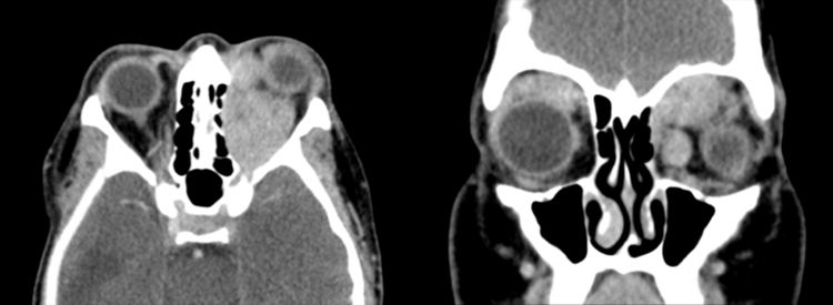
Computed Tomography (CT) of the orbits with contrast demonstrated bilateral orbital masses, markedly larger on the left, with marked left proptosis and encasement of the left optic nerve sheath—findings initially interpreted, in the context of her history, as most likely bilateral orbital metastasis from choriocarcinoma (Figure 2). CT and MRI scans of the brains otherwise showed no acute pathology and appeared stable from her prior ventriculoperitoneal shunt catheter placement.
Figure 2. CT orbits with axial and coronal cuts demonstrating bilateral masses, more significant on the left, with marked proptosis and complete encasement of left optic nerve sheath.
Staging CT of the chest, abdomen, and pelvis with contrast revealed multifocal systemic metastatic disease, including peritoneal and osseous (right inferior pubic ramus) metastases; left supraclavicular, axillary, subpectoral, and deep breast/chest wall lymphadenopathy; and bilateral pleural/subpleural soft-tissue nodules with rib erosions.
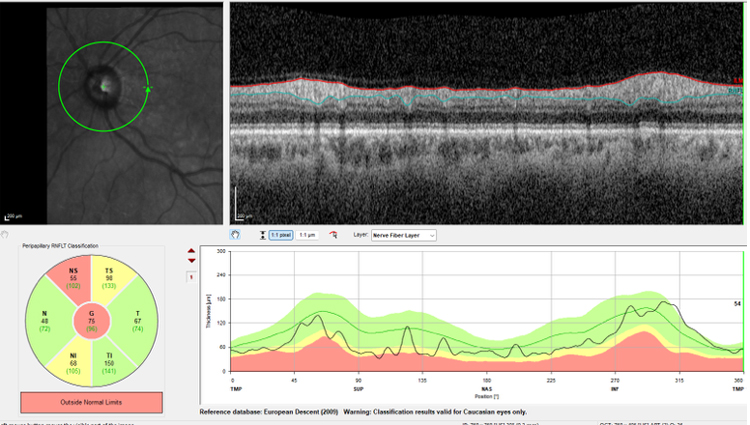
During inpatient admission, the patient's left visual acuity deteriorated precipitously overnight from hand-motion vision to no light perception, with OCT testing demonstrating further, now severe, nerve fiber layer thinning of the left eye (Figure 3). This prompted the decision to proceed with urgent orbital surgery for tissue diagnosis.
Figure 3. Optic nerve tomography of the left eye showing a significant increase in thinning superiorly, inferiorly, and centrally. At this point in the patient’s care, she no longer had light perception in her left eye.
She underwent a left anterior orbitotomy with orbital biopsy one day after initial presentation. Intraoperative findings were notable for anatomical disorganization with abnormal appearing indurated yellow/tan tissue.
Surgical pathology of the orbital lesions demonstrated high grade infiltrating predominantly epithelioid tumor cells that are positive for CK AE1/AE3, Mammaglobin, GATA-3, and CK7; and negative for synaptophysin, inhibin, hCG, p63, CD10, TTF-1, CK5 and SOX10. Altogether, the morphological and immunohistochemical profile was supportive of metastatic mammary carcinoma, lobular subtype with pleomorphic features—an unexpected result given the clinical–radiographic presumption of metastasis from her previous choriocarcinoma. This finding redirected the oncologic workup toward a breast/adnexal-type carcinoma primary and away from choriocarcinoma. Heme/oncology and breast oncology were engaged for systemic staging and biomarker-directed therapy, with PET-CT pending.
Postoperatively she was managed with topical antibiotic–steroid ointment to the incision, the corticosteroid taper, and aqueous suppression (ultimately dorzolamide-timolol) for persistent ocular hypertension. On serial follow-up, her left eye unfortunately remained without light perception, while the right eye remained functionally intact with stable nerve fiber layer and visual field, and she continued multidisciplinary oncologic care.
Discussion
This case reinforces that a new orbital mass in a patient with a prior malignancy must not be reflexively attributed to that cancer and emphasizes the importance of tissue diagnosis. The clinical syndrome—proptosis, motility restriction, and optic neuropathy and cross-sectional radiographic imaging are highly sensitive for the presence of an orbital process but poor for establishing histologic origin. A correct diagnosis is reached clinically or radiographically in fewer than half of orbital lesions in some series.[4] In our case, the radiographic impression favored choriocarcinoma metastasis on the basis of history, yet biopsy revealed a carcinoma of entirely different lineage, fundamentally changing the treatment pathway.
Breast carcinoma is the most common source of orbital metastasis, and its presentations are protean. Two well-described patterns exist: a mass-forming pattern producing exophthalmos, and an infiltrative, desmoplastic pattern—classically from invasive lobular carcinoma—that can paradoxically produce enophthalmos through fibrotic retraction of the globe.[1,6] Motility restriction and proptosis are the most frequent presenting signs, and bilateral involvement is well recognized.[3,7] Importantly, the histology of an orbital metastasis can be discordant with the known primary, which is precisely why obtaining a tissue biopsy is essential for proper management.[1]
Orbital biopsy is the diagnostic gold standard when histology will alter management. While many patients with a known primary and typical imaging are managed without biopsy, tissue sampling—via incisional orbitotomy, core-needle, or fine-needle aspiration—is essential when; the diagnosis is uncertain, the presumed primary is atypical for orbital spread, or lineage-specific and biomarker-directed therapy is contemplated.[3,5] In this patient, biopsy not only excluded the presumed choriocarcinoma metastasis but identified a lobular carcinoma whose receptor and androgen-receptor status carry direct therapeutic implications.
Management is multidisciplinary and generally palliative, aimed at preserving vision and quality of life. External-beam or stereotactic radiotherapy is the mainstay of local control for symptomatic orbital metastasis, achieving symptom improvement in the large majority of patients with an acceptable toxicity profile. Radiation therapy is usually combined with systemic therapy (chemotherapy, endocrine therapy, and CDK4/6 inhibition for hormone-receptor-positive breast carcinoma) according to tumor biology.[8-10] High-dose corticosteroids provide temporizing relief of compressive optic neuropathy, as employed acutely here. The prognosis of orbital metastasis is guarded—median survival is generally on the order of 1.5–2.5 years and reflects the burden of systemic disease rather than the orbital lesion itself, though metastasis from breast primaries carry comparatively favorable survival among orbital metastases.[1,3]
Finally, this case highlights the cost of lapsed cancer surveillance: an extended interval without oncologic and neuro-imaging follow-up allowed metastatic disease to advance to an irreversible, blinding orbital apex syndrome before detection. A high index of suspicion, prompt imaging, and early tissue diagnosis remain the cornerstones of care when orbital metastasis is suspected.
References
Sindoni A, Famà F, Vinciguerra P, et al. Orbital metastases from breast cancer: A single institution case series. Journal of Surgical Oncology. 2020;122(2):176–183.
Nghiem AZ, Islam M, Goh LY, et al. Presentation of Orbital Metastases. Ophthalmic Plastic and Reconstructive Surgery. 2025.
El-Hadad C, Koka K, Dong W, et al. Multidisciplinary Management of Orbital Metastasis and Survival Outcomes. Ophthalmic Plastic and Reconstructive Surgery. 2021.
Koukkoulli A, Pilling JD, Patatas K, et al. How accurate is the clinical and radiological evaluation of orbital lesions in comparison to surgical orbital biopsy? Eye (London). 2018;32(8):1329–1333.
Mombaerts I, Ramberg I, Coupland SE, Heegaard S. Diagnosis of orbital mass lesions: clinical, radiological, and pathological recommendations. Survey of Ophthalmology. 2019;64(6):741–756.
Homer N, Jakobiec FA, Stagner A, et al. Periocular breast carcinoma metastases: correlation of clinical, radiologic and histopathologic features. Clinical & Experimental Ophthalmology. 2017;45(6):606–617.
Yousef YA, Mohammad M, Khalil H, et al. Ocular and periocular metastasis in breast cancer: clinical characteristics, prognostic factors and treatment outcome. Cancers. 2024;16(8):1518.
Pierson TM, Tebit EV, El Sayed A, Smolkin ME, Dillon PM. Orbital metastases from breast cancer: retrospective analysis at an academic cancer center. The Breast Journal. 2016;22(4):404–411.
Pezzulla D, Di Franco R, Zamagni A, et al. Radiotherapy of orbital metastases: a systematic review of management and treatment outcomes on behalf of the Palliative Care Study Group of AIRO. British Journal of Radiology. 2023;96(1151):20230180.
Framarino-Dei-Malatesta M, Chiarito A, Bianciardi F, et al. Metastases to extraocular muscles from breast cancer: case report and up-to-date review of the literature. BMC Cancer. 2019;19(1):36.