Anterior Segment Optical Coherence Tomography: A Valuable Tool for the Anterior Segment Surgeon
Case report by Terrence Murphy, MD, MPH and Anjali Tannan, MD
Introduction
Optical Coherence Tomography (OCT) allows non-contact, high-resolution, cross-sectional imaging of ophthalmic tissues, aiding in diagnosis, treatment, surgical planning, research, and education. Initially used for evaluation of the retina and optic nerve, we are now finding more uses in the anterior segment. Anterior segment OCT (AS-OCT) provides high quality images of the cornea, anterior chamber, iris, angle structures, and the lens, without some of the limitations of ultrasound biomicroscopy. Reported uses of AS-OCT are ever-increasing, including evaluation of the ocular surface for neoplasias, corneal dystrophy management, post-op evaluation after endothelial keratoplasty, angle assessment during glaucoma workup, surgical planning for implantable collamer lenses, and many others.1 Here we present a use case of AS-OCT for diagnosis of a post-operative complication following cataract extraction and intraocular lens implantation.
Case Report
Our patient is a 72 year old right eye dominant male with a history of acute angle closure of the right eye, intermittent exotropia with facultative suppression of the left eye, who underwent urgent cataract extraction with intraocular lens implantation (CEIOL) of the right eye following multiple failed laser peripheral iridotimies. The patient’s CEIOL required use of a 6.25 Malyugin ring but was otherwise unremarkable. Four months after surgery, he was correctable to 20/30 and described his central vision as clear, but reported that his right eye’s vision was “brighter but distorted”. He described his vision as “like looking through a fish bowl or a glass of water”.
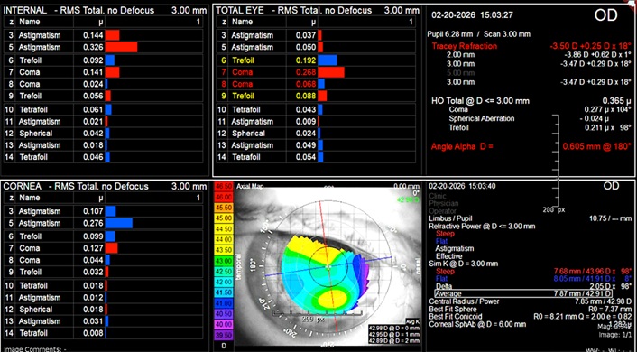
Manifest refraction and cycloplegic refraction with trial frames were conducted, without significant improvement in the patient’s visual experience. Slit lamp biomicroscopy was significant for a mid-dilated pupil, near-360 decrees of posterior synechiae, anterior pigment deposition, and possible tilt of the IOL. OCT of the macula was performed, ruling out any macular pathology. Tracey Technology iTrace was used to conduct aberrometry, identifying elevated levels of coma and trefoil, both corneal and internal.
Figure 1 I Trace aberrometry analysis of right eye
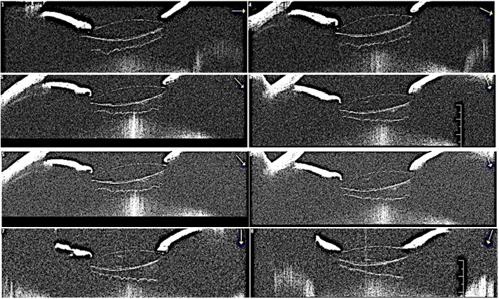
Anterior segment OCT was performed with results available below. The images demonstrated significant tilt of the intraocular implant both on the vertical and horizontal axes. Combined with the patient’s visual complaints and slit lamp findings, the patient was offered synechiolysis, intraocular implant exchange, and a pupillary cerclage.
Figure 2 Anterior segment OCT images. Arrows corresponding to the cross-sectional plane of the image
The patient was talked to the operating room. Synechiolysis was conducted without issue. During the implant exchange, significant inferotemporal zonular weakness was encountered during removal of the previous implant. A capsular tension ring and Ahmed segment were used to stabilize the capsule, and a B+L +32.0 Sofport L161AO was placed in the capsular bag. The lens appeared well-positioned and pupillary cerclage was conducted without issue.
The patient’s post-operative course has been complicated by slowly improving corneal edema. His pupil is approximately 3.5 – 4 mm in diameter and well-centered. He reports improvement in his symptom of asymmetric brightness, though visual acuity remains at 20/200 at post-op month one.
Discussion
Our case highlights an additional use for AS-OCT following CEIOL which allowed for the identification of sub-clinical IOL dislocation/malposition. In this scenario, we were highly suspicious of IOL malposition given the patient’s visual complaints, posterior synechiae, and faint IOL tilt. Here, the AS-OCT clinched the diagnosis, providing high-resolution images of the IOL’s tilted orientation in multiple planes. In retrospect, the AS-OCT findings also suggested a zonulopathy as a source of the lens’ malposition, that became apparent when encountered intraoperatively.
Examination by slit lamp biomicroscopy alone may not be enough for diagnosis and management of anterior segment abnormalities, even for the skilled user. AS-OCT allows for objective and high-fidelity examination of anterior segment pathology that may be too subtle to identify at the slit lamp. In pseudophakic patients specifically, this technology has been used to diagnose capsular bag distension, pseudophakic pupillary block, and uveitis-glaucoma-hyphema syndrome.2, 3, 4 Beyond generating helpful images, some software packages allow for measurement of size, shape, and relationship of the angle and other anterior segment structures. 5 More recently, AS-OCT is being integrated with surgical microscope systems, allowing for intraoperative evaluation of both the fundus and anterior structures. OCT has already proven to be a critical tool in the diagnosis and treatment of retinal pathology and glaucoma, and now also has a clear role in the diagnosis and treatment of anterior segment abnormalities.
References
Ramos, J.L.B., Li, Y. and Huang, D. (2009), Clinical and research applications of anterior segment optical coherence tomography – a review. Clinical & Experimental Ophthalmology, 37: 81-89. https://doi.org/10.1111/j.1442-9071.2008.01823.x
Tan, Y. L., Mohanram, L. S., Ti, S. E., Aung, T., & Perera, S. (2012). Imaging late capsular bag distension syndrome: an anterior segment optical coherence tomography study. Clinical Ophthalmology, 6, 1455–1458. https://doi.org/10.2147/OPTH.S34639
Mendrinos E, Dreifuss S, Dosso A, et al Evaluation of a pseudophakic pupillary block with an anterior segment OCT. British Journal of Ophthalmology 2008;92:714-715.
Lippera M, Nicolosi C, Vannozzi L, Bacherini D, Vicini G, Rizzo S, Virgili G, Giansanti F. The role of anterior segment optical coherence tomography in uveitis-glaucoma-hyphema syndrome. Eur J Ophthalmol. 2022 Jul;32(4):2211-2218. doi: 10.1177/11206721211063738. Epub 2021 Nov 29. PMID: 34841924.
Langenbucher, A., Szentmáry, N., Leydolt, C. et al. Calculation of ocular magnification in phakic and pseudophakic eyes based on anterior segment OCT data. Ophthalmic Physiol. Opt. 41, 831–841 (2021). https://doi.org/10.1111/opo.12822